Video dermatoscopy of lesions with computer analysis of malignancy

- Description of the examination
-
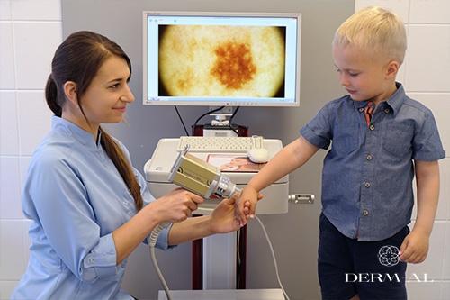
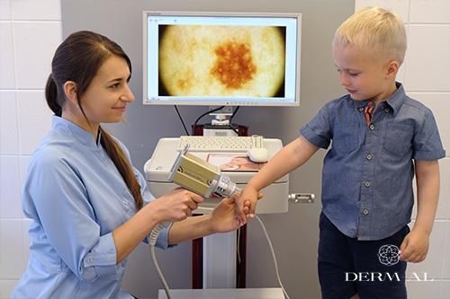
The Derm-Al Centre uses the advanced FotoFinder Vexia Gold system for digital dermatoscopy and medical photography documentation.
The video dermatoscope has a very sensitive digital camera that enables not only detailed analysis of respective lesions but also their evaluation “over time”. The device stores the history of examinations in order to compare changes in lesions between respective appointments. It is possible to take macroscopic pictures of the entire skin and identify if any new skin lesions developed since the last examination. During the examination, the physician and the patient can see lesions on the screen, and the physician advises the patient which of the lesions require specific attention.
Proper annual dermatoscopy as well as careful self-examination, i.e. observing if a lesion grows, changes its colour or structure, becomes painful or starts to itch or bleed, increase the chances of early detection of lesions that transform into untypical lesions or even melanoma, and help avoid unnecessary removal of benign lesions.
Video dermatoscopy with the FotoFinder Vexia Gold system enables:
- precise skin imaging with a magnification of 120x
- excellent quality photographs of skin lesions
- long-term storage of medical photographic documentation
- computer analysis of lesions for malignancy
- printouts for the patient
This way, it was possible to:
- diagnose early skin cancer
- control lesions and compare how they change
- detect even the most minute changes compared to the last examination
- store the results of examination
How does video dermatoscopy work
The physician takes pictures of the skin with the video camera in order to locate lesions. Then, he or she uses the digital dermatoscope built into the camera to take pictures of the lesions that the patient is concerned about or that are in the risk group, according to the physician. Then, the lesions are analysed by a computer program.
The photographs are catalogued and stored. If another examination is performed, the photograph of a lesion is compared to those from the previous examinations to identify changes, if any. The examination is painless and the patient can see the procedure on the screen, and he or she receives a printout of the examination report.Early diagnosis is the best protection against skin cancer!
Lesions must necessarily be examined by specialists if they have at least one of the following features:
- asymmetry, irregular shape
- blurred, jagged edges» irregular pigmentation - a lesion has different tones, e.g. dark brown, black and brown, reddish, grey and white
- more than 5 mm in diameter
- the shape changes: a lesion grows or becomes more protruding
-


